
Several areas of dying pines near Belleville, Illinois, were called to our attention in July, 1979 by Illinois extension advisor Mr. Wayne B. Siefert. An identical pine decline problem became epidemic in Urbana, Illinois during a prolonged summer drought in 1980. The disease symptoms were similar to those occasionally observed in pine plantations in localized areas for the past 25-30 years. The apparent cause for decline was initially diagnosed as a complex involving bluestain fungi and bark beetle attack during periods of drought stress.
Literature Review
Hartig reported, in 1878, the association between a blue-stain fungus, Ceratostomella pilifera, and bark borers that were affecting pine trees in Germany (5). In 1903, Von Schrenk reported a definite relationship between the infestation of pines by bark beetles and the development of a blue-staining fungus in lumber (12). Craighead also called attention to the constant association between “tree-killing” bark beetles (Dendroctonus) and blue-stain (4). A few years later, Rumbold reported investigations based on pine samples collected from dying shortleaf pines growing near Asheville, North Carolina (9, 10). The trees had been affected by a severe drought that occurred during 1925 and 1926. Many of the pines were infested with the Dendroctonus and Ips bark beetles, and the sapwood of these trees was affected by a fungus disease. Two blue-stain fungi were identified; Ceratostomella pini was associated with attacks of the insect Dendroctonus frontalis, and Ceratostomella ips was associated with a species of the Ips beetle.
In 1934, Steiner and Buhrer reported a pinewood nematode and named a new species Aphelenchoides xylophilus. It is possible that this nematode species may have been Bursaphelenchus xylophilus, the species presently associated with the pine wilt syndrome. The nematode apparently had become adapted to live in close association with blue-stain fungi and other woodinhabiting organisms in pine trees attacked by the Dendroctonus and Ips bark beetles (11).
Because of the economic interests of lumbermen, several researchers worked on the blue-stain fungi during the extensive drought of the 1930’s, when there was a substantial loss of pines in the southern part of the United States. Blue-staining greatly reduced the value of lumber cut from harvested logs. The affected pines deteriorated rapidly and were rendered worthless in one growing season (9). Apparently because the bark beetle population was high, trees injured by fire or heavy insect defoliation and those felled or injured by wind or by lumber operations were highly susceptible to bark beetle attack and the rapid development of blue-stain fungi.
An interesting history developed concerning the involvement of blue-stain fungi in the declining pine trees. Early pathological studies dealt with the symbiotic relationship of the fungus and various bark beetles. Limited work was reported concerning the potential pathogenicity of the bluestain fungi alone among various species of pines. Leach et al. (6) demonstrated that Ips bark beetles introduced blue-staining fungi and that the fungi are rarely introduced in any other way. They reported that C. ips occurs in the intestinal tract and on the bodies of the Ips beetle.
The blue-staining fungi have reportedly killed pitch pine (Pinus rigida) and shortleaf pine (P. echinata) (2, 3, 8). Mather (7) reported that C. ips and C. minor associated with the Ips and Dendroctonus bark beetles, respectively, may kill ponderosa pine (P. ponderosa). In extensive pathogenicity studies in 1970, Basham (1) demonstrated that C. ips, C. minor, C. montia (synonym: C. ips), and C. pilifera killed loblolly pine (P. taeda) and caused a deeply penetrating blue-stain of the sapwood. Two-year-old seedlings were more susceptible than 13-year-old sap-lings in terms of percentage of trees killed and average time to death. The pathogenic isolates penetrated living sapwood readily and caused blockage of the ascent of sap.
Many of the blue-stain fungi occurring in pine were originally classified in the genus Ceratostomella. These fungi are now placed in the genus Ceratocystis. Ceratocystis includes the prominent fungi that cause serious vascular diseases of trees. Ceratocystis ulmi causes Dutch elm disease, C. fagacearum causes oak wilt, and C. fimbriata causes canker-stain of London plane trees and stem-canker of quaking aspen. The internal vascular symptoms of canker-stain of London plane are similar to blue-stain associated with pine wilt. Fungi that have been reported as causal agents for blue-stain are C. allantopsora, C. ips, C. minor, C. minuta, and C. pilifera.
Symptoms
Initial symptoms of the pine blue-stain disease are observed when individual large branches start to exhibit either yellowing or rapid browning of needles. The branches may be either randomly distributed or they may occur predominantly on one side of the crown. Affected trees usually brown and die rapidly during the spring and summer (Fig. 1). The progression of symptom expression may extend over several weeks or months during the late fall and winter months. Trees exhibiting early branch symptoms during the winter may not completely die until warm spring temperatures occur.

An affected tree exhibiting advanced stages of the pine blue-stain disease in May 1981. At this stage extensive blue-staining in the wood of the trunk and branches can be observed. The pinewood nematode may also be present in many of the branches having needle browning.
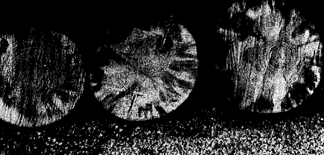
A blue-to-black discoloration of the sapwood develops in dying branches soon after the needles start to brown. The wood discoloration in cross-section is predominately wedge-shaped, being wide at the cambium and narrow in the inner sapwood (Fig. 2). Long sap streak discoloration develops predominately in the upper trunk and branches and extends downward into the lower trunk as the crown begins to die. In some cases where neighboring pine trees previously had been killed, new infected trees develop the blue streaking in the buttress roots and lower trunk, which may be indicative of root-graft transmission.

Wood discoloration in cross-section is wide in the sapwood and extends sometimes to the center of the affected trunk or branch. The individual blue streaks usually extend for several meters in the sapwood of trees exhibiting early symptoms of the blue-stain disease.
Scotch and Austrian pines appear to be the most susceptible species. Other pine species are susceptible but their relative susceptibility is not known.
Field and Laboratory Studies
The blue-stain fungus, Ceratocystis ips (Rumbold) C. Moreau, has been consistently isolated from Illinois pine trees exhibiting typical symptoms associated with the pine blue-stain syndrome. The fungus is widely distributed throughout most of Illinois. It has been isolated from infected wood of Austrian (P. nigra), red (P. resinosa), white (P. strobus), Scotch (P. sylvestris), Japanese red (P. densiflora), and Chinese red (P. tabulaeformis) pines. It has been consistently isolated from galleries of Ips grandicollis, the Southern pine engraver. Recently felled trees and slash are preferred breeding material for the insect, but the trunks, limbs, and small branches of trees under water stress are also subject to attack. In large living trees, the heaviest infestations are frequently found in limbs and the upper portions of trunks. During the drought of 1980, the Ips bark beetle was observed in large proportions, particularly among Scotch and Austrian pine growing in urban areas. The fungus C. ips has also been isolated from bodies of Monochamus carolinensis, the Carolina pine sawyer beetle1, which has been reported as a vector of the pinewood nematode. Ceratocystis species other than C. ips, have been isolated from dying pines, but taxonomic identifications are incomplete at present.
Ceratocystis ips isolations from dying pines were made from advanced margins of discolored wood, from synnema and ascocarp fruiting structures formed in insect galleries, and from the bodies of the two insect species mentioned above. The isolates grew rapidly on laboratory-prepared potato dextrose agar (PDA) at 24 C and develop typical colorless colonies that later turn dark gray to black after 7-10 days incubation.
Conidia were abundantly produced and were hyaline, 1-celled, clavate to ellipsoidal 1-2 × 3-10 μm. Synnemata were occasionally produced in culture and averaged 200 μm long. Ascocarps formed only when the fungus was grown on sterilized sections of pine wood placed on agar or on sterile, moistened filter paper in petri dishes.
Asci were never observed in the ascocarps. Ascospores were hyaline, 1-celled, oblong, surrounded by a hyaline gelatinous sheath that appeared rectangular (side view) with very short appendages or flanges at the ends of each spore (Fig. 3). They measured 1.5-2.0 × 3.0-4.0 μm. The ascocarps which formed superficially on the wood were black and approximately 190 μm in diameter with necks straight and sometimes bent and 560-900 μm long.

The ascospores of the Ceratocystis ips fungus have a characteristic gelatinous sheath and short flanges at the ends of the spores. (Enlargement of a picture taken through a phase microscope at 500× magnification.)
Pathogenicity Tests
Preliminary pathogenicity tests have been conducted with the fungus C. ips. Fifteen 2-year-old seedlings each of red, white, Scotch, and Austrian pine, growing in the greenhouse, were inoculated with spore suspensions. No external symptoms developed on any of the four species; however, internal vascular necrosis developed above and below the point of inoculation for distances of 1-5 cm in Scotch pine. The fungus was isolated from the margin of the necrotic areas 2 months after inoculation.
Scotch pines, 30 cm in diameter at breast height (dbh), were inoculated at several sites on the trunk and branches with three different isolates of C. ips on June 25, 1981. One of nine trees showed early branch symptoms 15 days after inoculation and was 60 percent dead 6 weeks after inoculation. The fungus was recovered from trunk samples and from branches showing needle symptoms. The pinewood nematode, Bursaphelenchus xylophilus, was also recovered from several branches showing advanced browning of the needles. Three other inoculated trees exhibited branch symptoms 45 days after inoculation and C. ips was isolated from the trunk and from those branches exhibiting needle browning. Pinewood nematodes were not recovered from the affected branches or from trunk samples of these trees.
Discussion
During most of the 1981 growing season, rainfall was above average and the field-inoculated trees in my test were never under water stress. Evidence indicates that the pine blue-stain disease increases during serious drought years and for one or more years following the drought. In central Illinois, the disease incidence appeared to have been lower during 1981 than in the drought year of 1980. Drought may be one of the most important factors predisposing susceptible pine species to bark beetle invasion and the subsequent infection of living trees by blue-stain fungi.
In previous pathogenicity trials on large pines, the isolates of blue-stain fungi killed inoculated pines only when the investigator used multiple trunk or branch inoculations. Among naturally-infected trees, numerous individual, long, wedgeshaped, blue-stained areas are usually observed around the circumference of sectioned trunks and branches (Fig. 2). Each stained area appears to represent individual points of inoculation by bark beetles. Extensive areas of the cambium are girdled by initial bark beetle invasion of the upper trunk and branches. Immediately following beetle inoculation, the blue-stain fungi grow rapidly, both vertically and radially, through the outer vascular tracheids and ray parenchyma. Large random areas of the vascular system become inoperative and the affected tree becomes highly attractive to additional beetle attack.
The pinewood nematode is frequently cited as the primary pathogen of pine wilt, but I am not certain of its involvement, either as a single or primary pathogenic agent in the death of pine trees in Illinois. It is apparent that a complex relationship exists between various species of bark beetles, the blue-stain fungi, and the pinewood nematode. Some entomologists indicate that bark beetles are vectors of the pinewood nematode. Most of the insect species reported to carry the nematode also are reported to transmit blue-stain fungi. The Ips bark beetle is believed to be a primary vector of the blue-stain fungus in Illinois.
The “pine wilt disease,” as it is popularly called, may be caused by one pathogenic agent or a combination of pathogenic agents and environmental factors, depending upon where the pines are dying in the United States. Pine wilt presently refers to dying pine trees infested with the pine wilt nematode and not to the actual symptoms of wilt. Confusion will occur indefinitely in the literature unless more specific names are used for the various infectious and noninfectious diseases that cause decline and death of pine trees.
After reviewing the current literature on the pine wilt disease, it was apparent that the blue-stain fungi curiously have been regarded as saprophytic or secondary agents in the disease complex. Although blue-stain fungi may be weakly pathogenic, the virulence of various species and strains needs extensive testing on a wide range of pine species to determine if the pine wilt syndrome is primarily caused by the pine blue-stain disease.
Footnotes
↵1 The Ips beetle was identified by John K. Bouseman and the Carolina pine sawyer beetle was collected by James E. Appleby, both entomologists at the Illinois Natural History Survey.
- © 1982, International Society of Arboriculture. All rights reserved.
In this issue


Jump to section
Related Articles
Cited By...
- No citing articles found.