
Abstract
Although the presence of bleeding cankers on deciduous trees may raise fears of the presence of Phytophthora ramorum, the cause of sudden oak death, other pathogens also cause similar symptoms. This review of hardwood tree diseases with bleeding canker symptoms provides an overview of available information of these diseases, providing a diagnosis guide as well as a stimulus for continued research in these areas.
Sudden oak death, caused by Phytophthora ramorum, has become a serious concern in both the forest and nursery industries since the disease was first discovered in California, U.S. in 1995 (Henricot and Prior 2004). The pathogen is relatively new to North America and has a broad host range that includes species in at least 50 genera of Gymnosperms and Angiosperms (Rizzo et al. 2005). Symptom expression of the disease on oaks is often described as lesions on the main stem where fluid is oozing through bark fissures or where stains on the bark suggest that such fluid was recently present. Tissues beneath the stained areas are brown and no longer functional (Rizzo et al. 2002). These necrotic areas are called “bleeding cankers” and their presence on trees anywhere now raises fears that P. ramorum may have spread beyond the West Coast of the United States. However, because many other pathogens cause bleeding on a variety of hosts, diagnosis should be approached with an open mind. Bleeding, oozing, or stained bark can result from infection by fungal or bacterial pathogens as well as injury caused by insects, mechanical wounding, or cambial death in stressed trees.
Determining the cause of a bleeding canker can be a difficult task. If the cause is either P. ramorum or one of the many other species of Phytophthora, then a kit containing antibodies from mammalian immune systems coupled with specialized enzymes to detect large molecules unique to species of Phytophthora can be useful. Unfortunately, the kits do not enable determination of which species of Phytophthora is present. That requires culturing of the pathogen on laboratory media, examination of reproductive structures and culture morphology, and—perhaps—analysis of DNA sequences.
If preliminary tests suggest that Phytophthora is not a factor, the diagnostic process can become more complex, and both prognoses and management strategies can be more difficult to find. Reports of bleeding cankers in the literature often include little if any information about the causal organisms and nothing about management strategies. The following literature review of tree disease characterized by the presence of bleeding cankers is an effort to consolidate many published reports in a way that will provide an overview of our current state of knowledge, aid diagnosis, and stimulate further research.
MAPLE (ACER)
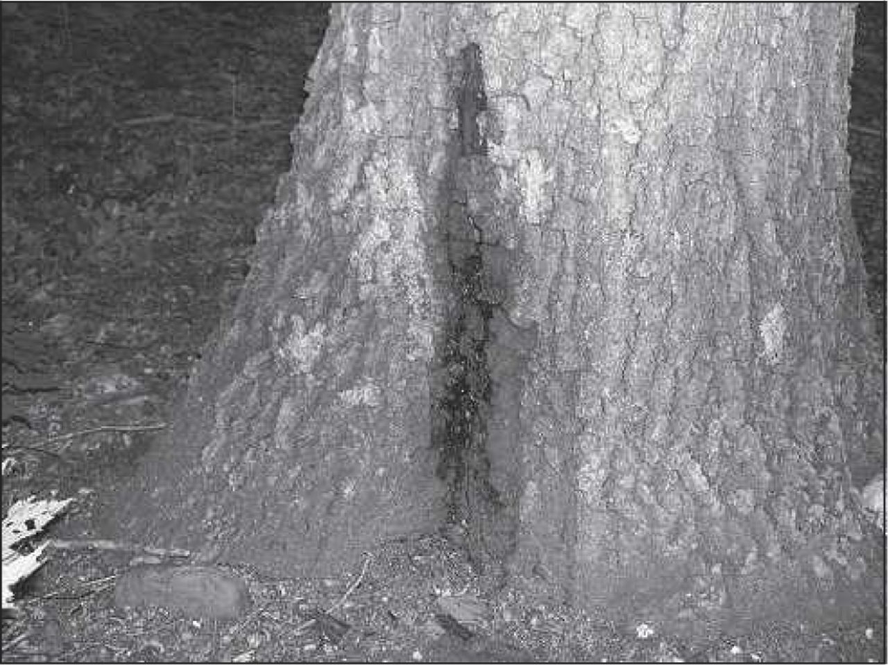
Phytophthora cactorum causes bleeding cankers on many species of maples with symptoms described as indefinite cankered areas that ooze reddish brown fluid in the fall (Caroselli and Howard 1940; Caroselli 1953). Discoloration extends into the sapwood beneath the cankered area and is outlined by an olive-green margin. The necrotic area can contain a reddish brown watery fluid that may expel forcefully when punctured. A general decline of the tree is also often observed. Incidence of bleeding cankers on maples may increase when trees experience stress as a result of heavy salting (Lacasse and Rich 1963). In addition to P. cactorum, similar symptoms on maple caused by P. cambivora, P. cinnamomi, P. citricola, and P. palmivora have been reported (Figure 1) (Drillias et al. 1982; Barnard and Mitchell 1993; Erwin and Ribeiro 1996). These reports are the result of consistent isolation of these pathogens from the cankers and not from inoculation experiments. Some trees are able to successfully compartmentalize the infection if it is limited (Caroselli 1953). Caroselli (1953) also notes that removal of infected tissue has not been a successful control strategy. Fungicides may be effective but are not often economically feasible (Erwin and Ribeiro 1996).

Typical bleeding canker symptoms caused by Phytophthora species on sugar maple.
Unique Features of Phytophthora
Microbes in the genus Phytophthora used to be classified as fungi until recent DNA analysis proved that they are actually more closely related to brown algae and are best included in the Kingdom STRAMINIPILA. All species produce asexual zoospores with flagella that allow them to swim short distances in free water. They also reproduce sexually through formation of thick-walled oospores. Most species of Phytophthora live in the soil where they parasitize plant roots, but a few—such as the Sudden Oak Death pathogen and P. infestans, cause of the Irish potato famine—thrive above ground when rainfall is abundant. Phytophthora spores do not survive for long when exposed to sun or dry air; they are more likely transported long distances in infested soil or plant material. In any case, excess moisture is a key element in triggering a Phytophthora-caused plant disease outbreak.
Bleeding cankers caused by Fusarium solani were reported on sugar and red maple by Wood and Skelly (1964). Initial black exudates are followed by the development of sunken areas and eventually the bark breaks away. Cankers developed when isolates of the pathogen were inoculated into healthy maples (Wood and Skelly 1964). Inoculations conducted during the spring produced black exudates by the next fall (Skelly and Wood 1966). Weidensaul (1968) found that F. solani was easily cultured from the bark of healthy sugar maples and hypothesized that any wound could be the site of introduction of the pathogen to the inner bark. If wounding occurs during the dormant season, then a canker develops. Once the tree resumes growth in the spring, it is able to contain the pathogen and eventually close over the canker (Wood and Skelly 1969).
Drillias et al. (1982) describe a basal canker of sugar maple caused by an unidentified Fusarium species. Seemingly healthy bark at the base of the trunk exudes sap and covers necrotic, dark reddish brown tissue that extends into the wood. A Fusarium spp. was consistently isolated from bark and wood chips taken from the margins of these cankers. Wound inoculations of healthy trees produced similar symptoms.
Cryptosporiopsis canker, caused by an unidentified species of Cryptosporiopsis, appears as brownish black exudates at the point of infection on red maple saplings (Taylor and Moore 1979). Phloem and cambium underlying the symptomatic area are discolored in an elongated, tapering shape. Taylor (1983) was able to reproduce symptoms with inoculations performed by drilling holes into the tree and inserting colonized agar plugs. The presence of tree cricket eggs in holes at the center of cankers prompted further investigation into the possibility of an insect vector of the pathogen. Taylor (1983) showed that crickets exposed to cultures of Cryptosporiopsis spp. could successfully transfer the fungus to a new agar plate. However, crickets exposed to the fungus and then transferred to a maple branch induced cankers around oviposition holes in only a few instances. Nevertheless, the disease is generally considered to be spread by the narrow-winged tree cricket (Sinclair and Lyon 2005).
HORSECHESTNUT (AESCULUS)
Bleeding bark lesions develop on horsechestnut when the tree is infected by Phytophthora cactorum or P. citricola. These pathogens cause crown dieback accompanied by rusty red fluid flowing from patches of dead bark on the trunk. The exudate then dries to form a black crust (Brasier and Strouts 1976). In this same report, plugs of agar colonized by either P. citricola or P. cactorum were inserted under the bark of healthy horsechestnuts. Within 3 months, bleeding regions were observed at the point of inoculation. The pathogen was successfully reisolated from the new lesions. Removal of branches with cankers may prevent spread to the rest of the tree (Brasier and Strouts 1976), and Webber (2005) also suggests that excision of small cankers from the trunk may be a successful disease management strategy.
Bacteria have been frequently isolated from bleeding cankers on horsechestnut, leading to the assumption that the bacteria cause the symptoms. However, the bacteria have not been identified, and no inoculation studies have been performed (Sinclair and Lyon 2005).
MADRONE (ARBUTUS)
Phytophthora cactorum was identified as the cause of bleeding cankers on Madrone in Washington and California (Stuntz and Seliskar 1943; Wagener and Cave 1944). Watersoaked areas of bark cover discolored tissue. Eventually the dead bark sloughs off. Cracks produced during this process often exude a dark liquid consisting of decomposed host material. Trunk symptoms are accompanied by crown dieback and general decline of the tree. Stuntz and Seliskar (1943) inoculated healthy mature Madrone by placing mycelium of P. cactorum underneath the bark of stems and roots. Cankers developed in both the stem and the roots and the pathogen was reisolated from the infected tissue. When cankers are small, excision may be successful in halting their growth (Stuntz and Seliskar 1943).
Madrone is also a host of P. ramorum and will exhibit bleeding cankers when the trunk is infected. Infected trees have been found in California (UK PRA 2003).
BIRCH (BETULA)
Although both Howard (1942) and Caroselli (1953) report Phytophthora cactorum as a pathogen of birch, there is little else describing the associated disease. A general decline of the tree is accompanied by cankered areas of the trunk and branches, which exude reddish brown liquid.
CHESTNUT (CASTANEA)
A recent surge in the incidence of ink disease in Europe and the United States has renewed interest in this disease (Crandall et al. 1945; Vettraino et al. 2001). Infection by one of two pathogens, P. cambivora or P. cinnamomi, begins in the roots as light brown and green lesions and progresses to produce symptomatic wilting and dieback of the crown. Blue–black exudates from the root lesions stain the soil and give the disease its name. Occasionally, lesions extend up the trunk above the soil line (Crandall et al. 1945). Crandall et al. (1945) performed inoculation experiments by repotting healthy seedlings into soil from which infected seedlings had been removed. This work showed American and sweet chestnut species to be highly susceptible. Day (1939) conducted inoculation experiments by placing mycelium into a slit made into the bark of sweet chestnut. Cankers developed, although it is noted that in field conditions, a wound is seldom seen in conjunction with the development of a canker. Day (1938) found that ink disease incidence was much higher in poorly drained soils. If only small portions of the roots are infected, he suggested that clearing away all soil and leaving it exposed may prevent spread within the tree. Diseased bark can also be excised (Day 1939).
CITRUS (CITRUS)
Several species of Phytophthora can infect citrus and cause symptoms of necrotic bark oozing gum. The inner bark becomes discolored and infused with gum, which then oozes out. Fawcett (1923) describes the gum as clear and watery, originating from the wood, although decay is limited to the bark. This disease is called foot rot, Phytophthora gummosis, brown-rot gummosis, collar rot, and mal di gomma (Knorr 1973). The amount of exudation is dependent on the host species and weather conditions (Klotz 1973). Phytophthora citrophthora, P. parasitica, and P. nicotianae have all been shown to cause these symptoms (Knorr 1973). Noninfested plant material planted in well-drained soil is usually an effective management strategy. There are also resistant plants and fungicide-treated seed available. Once infection occurs, fungicides can be used or infected tissue can be excised (Knorr 1973).
DOGWOOD (CORNUS)
Phytophthora crown rot of dogwood is caused by P. cactorum and has been reported from the New England, U.S. area and Washington State (Horst 2001). Lesions at first are inconspicuous, although removing the bark may reveal brown necrotic areas. As the cankers develop, the bark breaks and fluid oozes out. Crown symptoms also develop (Creager 1937). Pathogenicity was confirmed with dogwood seedlings and larger trees in the field by wounding the stem and inoculating with mycelium (Creager 1937; Stuntz and Seliskar 1943). Stripping away infected bark can halt canker advancement because the pathogen is sensitive to dryness and heat (Stuntz and Seliskar 1943).
RUSSIAN-OLIVE (ELEAGNUS)
Phytophthora cactorum has been reported from Russian-olive. When isolates from a bleeding canker on Russian-olive in Illinois were inoculated into greenhouse-grown trees, new cankers formed but reisolations from the cankers were only successful within 6 months of inoculation (Carter 1953).
BEECH (FAGUS)
Day (1932) reports P. cambivora as a pathogen of European beech and describes the disease as similar to, although less virulent than, the ink disease of chestnut. Wilting and yellowing of the crown develops in conjunction with dead areas of bark near the base of the trunk, which exude a clear fluid that later darkens (Day 1938). Inoculation experiments showed European beech is highly susceptible to P. cambivora, but not P. cinnamomi, another pathogen associated with ink disease. Beech saplings were inoculated by inserting agar plugs into bark slits made at the base of the trunk (Day 1939). P. cambivora attacks the roots of trees under high moisture conditions; promoting root health and maintaining proper drainage may control this disease (Day 1939).
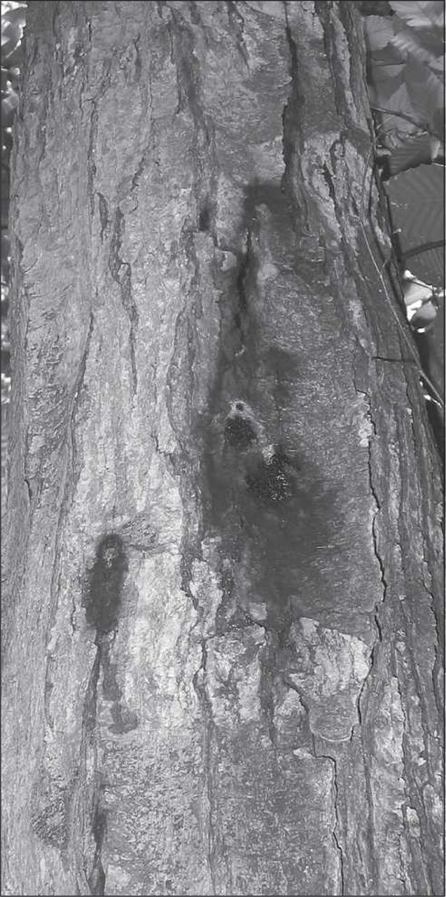
Caroselli (1953) and Howard (1942) report P. cactorum as a pathogen of American and European beech. Recent surveys of declining European beech in the northeast United States have found P. cactorum associated with some cankers. These same surveys have found that members of the Phytophthora citricola species complex are more frequently associated with bleeding cankers on European beech (Nelson et al. 2006). In both cases, black patches of bark are accompanied by rusty brown exudation; necrosis extends into the sapwood (Figure 2). Field observations of some successfully compartmentalized cankers suggest that environmental predisposition may favor this disease and has led to the recommendation of management through maintaining tree vigor and avoiding soil compaction (Jung et al. 2005).

Bleeding canker of European beech caused by Phytophthora citricola.
WALNUT (JUGLANS)
A bacterium, Brenneria rubifaciens, causes deep bark canker of walnut. First discovered in California in the 1950s, this disease is now also found in Europe (Wilson et al. 1967; Gonzalez et al. 2002). Dark longitudinal streaks run along the inner bark and outer sapwood. Cracks in the bark develop and a slimy substance is exuded, especially in the summer (Wilson et al. 1967). In Europe, B. rubifaciens was inoculated into deep wounds made in the trunk. Two months later, inner bark necrosis was found, but it took 4 years for external exudation to be produced. The bacterium was reisolated from the canker and from the exudates (Gonzalez et al. 2002). Wilson et al. (1967) also performed a similar inoculation study with the same results, although cankers produced the slimy exudates in as little as 6 weeks.
A closely related bacterium, Brenneria nigrifluens, causes shallow bark canker of walnut. Discolored spots on the bark of the trunk or branches exude dark brown fluid that spreads out over the bark during the summer. The discoloration usually is contained in the outer bark, but occasionally pockets of necrotic tissue extend to the cambium (Saccardi et al. 1998). Cankers several years old sometimes have a ring of callus tissue surrounding them (Morone et al. 1998). Wilson et al. (1957) inoculated wounds; exudation developed in 4 weeks, and the bacterium was reisolated from the canker and from the black exudates. This disease is a problem in California, southern Europe, and the Middle East (Sinclair and Lyon 2005).
Although these two diseases have similar symptoms and a similar geographic distribution, deep bark canker is more serious because it can penetrate to the cambium and therefore has a higher mortality (Holdeman 1970). Because shallow bark canker does not significantly decrease yield or harm the tree, there are no published control measures. Deep bark canker can be avoided by reducing stress and by sanitizing any harvesting or pruning equipment used on the tree. Excision of diseased tissue does not control the disease; apparently, the bacterium resides in asymptomatic tissue far from the cankers (Teviotdale et al. 2002).
SWEETGUM (LIQUIDAMBAR)
Three reports of outbreaks of bleeding canker symptoms on sweetgum indicate that Botryosphaeria ribis is the causal agent, but differences in symptoms and geography make understanding of this disease difficult.
Pirone (1942) first described a bleeding necrosis of sweetgum in the New York City, and New Jersey region. A heavy motor oil-like substance bled from apparently unwounded areas of bark. Inner bark was dark reddish brown and, in the advanced stages, this discoloration extended into the cambium. Pockets of a white crystalline solid were sometimes found within the necrotic bark. Crown decline was common but no root symptoms were seen. In this study, no inoculation experiments were performed, but fruiting bodies of Botryosphaeria ribis were observed on the bark.
In 1957, Toole and Morris (1959) observed a trunk lesion on sweetgum in the Gulf Coast Region of the United States. Lesions began as gum oozing from the bark. The gum flowed down the trunk where it turned black and hardened. Callus tissue forming around the cankered area created ridges under the bark, and the inner bark and cambium were discolored. The fungus B. ribis was frequently isolated from diseased tissue, and cankers developed when mycelium was inserted into slits cut in the bark and cambium of mature sweetgum (Toole and Morris 1959; Toole 1963).
Neely (1968) described a bleeding necrosis of sweetgum in Illinois and Indiana. Bleeding cankers were accompanied by an unpleasant sweet odor. Multiple cankers on one tree were interconnected by a reddish brown discoloration in the sapwood. Isolations from the sapwood and from the fruiting bodies on the bark produced cultures of B. ribis. Inoculations using colonized agar plugs placed into chisel wounds in the sapwood in the fall produced characteristic cankers by the next spring and bleeding by the summer.
Although all of these studies concluded that B. ribis was the causal agent, the differences in geographic location and symptom description make it difficult to ascertain if this is the same disease. Although Pirone (1942) reports that the disease is often rapidly lethal, authors of other reports do not report the fate of the trees.
YELLOW-POPLAR (LIRIODENDRON)
In 1956, decline of yellow-poplar led to the discovery of a canker disease caused by Fusarium solani (Dochinger and Seliskar 1962). The initial symptoms are difficult to detect. Small cracks appear in the bark, but the bark remains on the tree although it is necrotic. Later, lesions begin to ooze, usually during the fall as the fungus becomes active. Wounding seems to be necessary for disease (Dochinger and Seliskar 1962). Anderson and Hoffard (1978) found all cankers in one region in Ohio had ambrosia beetle injury in the center, providing one possible mode of fungal transmission. Dochinger and Seliskar (1962) performed inoculations by cutting out the bark to the cambium, inserting a disk of colonized agar, and then replacing the bark. Cankers developed and grew for 1 year but became inactive after that as callus tissue built up and the tree contained the infection. Further inoculation tests showed that inoculations made in the spring and summer were inactive by the next year, but inoculations made in fall produced cankers still active 2 years later (Dochinger and Seliskar 1962).
TANOAK (LITHOCARPUS)
Tanoak (Lithocarpus densiflorus) is highly susceptible to sudden oak death caused by P. ramorum. Bark turns black where the pathogen is active, and dark red sap oozes from fissures in those areas. After the cankers appear, the crown of an infected tree wilts. Rizzo et al. (2002) confirmed pathogenicity by inoculating tanoak seedlings and mature trees. Mycelium was inserted into slits or bore holes in the bark above the soil line. Both methods were successful in reproducing symptoms; mature trees began to ooze sap 3 weeks after inoculation. Because sudden oak death and Phytophthora ramorum are newly discovered issues in plant health, management strategies are limited. Phosphites have been approved for treatment of infected trees through direct injection or applied as a bark drench coupled with surfactant–penetrants (Rizzo et al. 2005). Control measures are also focused on prevention of spread; quarantine regulations are in place to prevent the spread of the pathogen from the West Coast (Rizzo et al. 2005).
APPLE (MALUS)
Botryosphaeria canker emerged as an important problem of apples in 1952 in Indiana and now causes tree loss from New York to Georgia. The pathogen, Botryosphaeria dothidea, is able to take advantage of drought and cold-stressed trees and is especially common around frost cracks and pruning wounds. The cankers begin as small sunken reddish lesions, which then form blisters. These blisters can crack and exude a watery substance. Eventually the necrotic areas expand to form an elliptic canker that may girdle the stem, but most trees are able to compartmentalize effectively (Brown and Britton 1986).
AVOCADO (PERSEA)
Although Phytophthora heveae has been found from soil samples in Tennessee and North Carolina, bleeding canker on avocado caused by P. heveae has only been found in Guatemala (Campbell and Gallegly 1965; Zentmyer et al. 1978). Trees show symptoms of cracking and bleeding on the lower trunk and upper roots. The inner bark has brown necrotic tissue with a distinct margin; the wood is rarely infected. Isolates of P. heveae inoculated into the stems of avocado seedlings produced lesions (Zentmyer et al. 1978).
POPLAR (POPULUS)
Cryptosphaeria canker on poplar is caused by Cryptosphaeria populina (syn. C. lignyota). Infection begins in the wood and then spreads to the cambium and bark. Symptoms first appear as infected bark becomes discolored; then bleeding occurs at the canker margins, where a reddish brown fluid exudes from the bark (Hinds 1981; Sinclair and Lyon 2005). Cryptosphaeria canker is widespread throughout the range of aspen, particular in the western United States (Hinds 1981). Although Cryptosphaeria canker is not the most common canker disease of poplar, it does have a high mortality rate because it frequently infects younger trees, which are more easily girdled. Juzwik et al. (1978) surveyed trees in Colorado and attributed one-fourth of tree mortality to Cryptosphaeria canker.
Ceratocystis fimbriata causes cankers on poplar with symptoms of bleeding cankers. For the fungus to infect the tree, a wound is required (Manion and French 1967). The fungus is carried to fresh wounds by various insect vectors, most commonly nitidulid beetles (Hinds 1972b). The elliptic canker develops as a depression in the bark around the wound. Often the face of the canker becomes covered with amber-colored sap exuding from the margins, and rusty brown fluid can also bleed from the margins (Hinds 1972a; Sinclair and Lyon 2005). Growth halts after the first dormant season, but fruiting bodies develop on the canker and underneath the bark after two growing seasons and cankers are apparent for several more growing seasons (Manion and French 1967). Cankers developed when Wood and French (1963) inoculated C. fimbriata into wounds on the main stem. This disease is known throughout the range of aspen but is more common when site conditions are poor (Manion and French 1967; Hinds 1972a).
STONE FRUIT (PRUNUS)
Bacterial cankers caused by Pseudomonas syringae are light tan with a water-soaked appearance, a sour odor, and gummosis (Hepting 1971; Weaver 1978). Both the sapwood and cambium show signs of necrosis (Klement et al. 1984). Many important fruit trees, including members of the genera Prunus and Pyrus, are hosts (Hepting 1971). Girdling by the cankers on the branches decreases productivity and may kill the tree.
The bacterium relies on cold temperatures to cause symptoms. It reduces the sugar content of the tree and has ice-nucleating proteins to promote frost damage and susceptibility. When temperatures dip low enough, the bacterium is able to promote the formation of frost, causing the symptoms to appear (Klement et al. 1984). Therefore, plants can have high concentrations of bacteria but be asymptomatic (Cameron 1970). Because of this, treatment is difficult. Cameron (1970) showed that the bacterium was found far from symptomatic tissue. Consequently, removal of infected branches may not be effective. Bactericide sprays may be effective if infection is not yet systemic, but this is difficult to determine (Cameron 1970).
Pruning wounds infected by Phytophthora syringae can lead to oozing cankers in almond. Bostock and Doster (1984) used isolates collected from cankers to inoculate pruning wounds; cankers similar to the naturally infected trees developed (Bostock and Doster 1984). Wounds are necessary for P. syringae to infect the tree. The treatment of wounds with fungicides, or the avoiding of wounding during the cooler months, when the pathogen is active, may prevent this disease (Ogawa and English 1991).
OAK (QUERCUS)
Phytophthora ramorum, although first identified as the cause of blight on rhododendron and viburnum in Europe, is now rapidly gaining notoriety as the cause of sudden oak death in California and Oregon. P. ramorum is pathogenic on a wide range of plants. Oak species infected include Q. kelloggii, Q. parvula, Q. agrifolia, and Q. chrysolepsis in California and Q. cerris, Q. falcata, Q. ilex, and Q. rubra in Europe (Henricot and Prior 2004). Inoculation experiments conducted on detached logs indicate the host range may actually be much broader (UK PRA 2003). Bleeding cankers develop as brown–black patches of bark seep dark red sap. This is combined with severe wilt and dieback of the crown and eventual death of the tree (Rizzo et al. 2002). Rizzo et al. (2002) inoculated mature trees with mycelial plugs placed into holes made with a cork borer. The bark was then replaced, and cankers developed on all inoculated trees within 6 months.
Because of the recent discovery of this pathogen and the diseases it causes, disease management strategies are still being developed. In Europe and in Oregon, efforts have focused on eradication, whereas in some areas of California, the pathogen is considered well established, necessitating management toward quarantine efforts (Henricot and Prior 2004). Once an area has become infected, management strategies depend on the intended scale of the treatment. Single trees on the interface of the urban and wildland are often highly valued by homeowners. Phosphite fungicides are available for trunk injection and can be also be applied with a surfactant as a bark drench. On the national level, control is focused on restricting interstate movement of potentially infected plants. Treatment on the forest landscape level is the most difficult given the broad host range of the pathogen and because little is known of its biology and ecology (Rizzo et al. 2005).
Surveys in California for P. ramorum have revealed P. nemorosa is also associated with bleeding cankers on tanoak and coast live oak (Hansen et al. 2003). P. nemorosa occurs sporadically and does not seem to spread as rapidly or cause as much mortality as P. ramorum (Rizzo et al. 2005).
Miller (1941) identified bleeding cankers on coast live oak in California, isolated P. cactorum, and inoculated healthy oaks to produce identical symptoms: thin light brown to thick reddish exudates from bark fissures dry to form black tar-like areas on the surface of the bark. Howard (1942) documented the same disease in the Northeast. Mircetich et al. (1977) performed inoculations with P. cactorum, P. cinnamomi, and P. citricola to successfully reproduce similar symptoms on oak (Figure 3).
Bleeding canker on oak typical of symptoms caused by Phytophthora species (photo courtesy of Y. Balci).
In 1953, a severe drought in West Virginia created stressful conditions. True and Tryon (1956) documented an outbreak of bleeding cankers on oaks after the drought. Crown dieback and leaf discoloration was often accompanied by bleeding cankers on the trunk. Isolations from cankers did not result in any dominant putative pathogen; 89% were classified as sterile. Exploration into the factors related to cankering showed a high correlation between canker incidence and dry conditions on high ground (True and Tryon 1956).
Bleeding cankers on oak associated with unidentified bacteria have been reported. These cankers are sometimes associated with insect wounds. Necrosis extends from the outer sapwood to the surface of the trunk and exudates stain the surrounding bark (Sinclair and Lyon 2005).
WILLOW (SALIX)
Erwin and Ribeiro (1996) and Caroselli (1953) list willow as a host of Phytophthora cactorum with symptoms of bleeding cankers.
LINDEN (TILIA)
Erwin and Ribeiro (1996) and Caroselli (1953) list linden as a host of Phytophthora cactorum with symptoms of bleeding cankers.
ELM (ULMUS)
Howard (1942) lists elm as one of the trees susceptible to Phytophthora cactorum with symptoms of bleeding cankers.
BEYOND NORTH AMERICA
Many other pathogens capable of causing bleeding canker symptoms are common on tree species found in North America. However, the cankers themselves have not been found in North America. For instance, Webber (2005) notes bleeding cankers on linden in England caused by P. citricola and P. cactorum, but these host–pathogen combinations have not been found in North America. Bleeding cankers on European beech in Europe have been shown to be associated with at least six species of Phytophthora, although P. citricola was the most common (Jung et al. 2005). Cankers on beech in Italy were shown to be caused by P. pseudosyringae, which had not previously been recorded (Motta and Annesi 2003). In addition, P. ramorum has been isolated from bleeding cankers on European beech, horsechestnut, and ash, and log inoculation tests prove pathogenicity (UK PRA 2003; Rizzo et al. 2005). Log inoculations have shown sweet chestnut to be susceptible to P. ramorum, but only leaf symptoms on chestnut have been found naturally occurring in England (UK PRA 2003; Denman et al. 2005). The newly described Phytophthora kernoviae (formerly Phytopthora taxon C) has been found associated with bleeding cankers on European beech, English oak, and yellow-poplar (Brasier et al. 2005). Both P. ramorum and P. kernoviae have been isolated from adjacent cankers on the same tree, which raises fears of the possibility of hybridization (Brasier et al. 2004). A hybrid Phytophthora has been shown to cause bleeding cankers and decline of alder in Europe (Figure 4) (Brasier et al. 1995). The bacteria Brenneria alni on alder and Brenneria quercina on oak cause bleeding cankers (Hildebrand and Schroth 1967; Surico et al. 1996).

Bleeding canker on alder caused by unidentified Phytophthora species.
ADDITIONAL CONSIDERATIONS
Although the presence of bleeding cankers may signify the presence of a pathogen, bleeding can also be the result of mechanical or insect damage. Some trees are more prone to extensive sap flow after pruning, whereas others may produce and exude more fluids in response to insect activity. For example, the dried black exudates of Fusarium canker on red maple may look similar to insect injury (Skelly and Wood 1966). Russian-olive may exhibit bleeding resulting from insects, and if insect tunneling is evident under the bark, this is a potential cause (Riffle and Peterson 1986). Especially in the spring, when trees may have larger vessels near the surface of the bark and are moving large amounts of fluids as a result of increased biological activity, any sort of wounding, whether by insects, pruning, or other mechanical damage, may result in the tree exuding or bleeding sap.
IMPLICATIONS FOR ARBORICULTURE
Based on this summary of diseases of hardwood trees with bleeding cankers, it is apparent that many reports of such a condition exist without further research to establish the etiology of the disease. The increased focus on bleeding cankers brought on by the surge in lethal diseases with this symptom will hopefully lead to an increase in research and subsequent understanding of these diseases.
- © 2007, International Society of Arboriculture. All rights reserved.
LITERATURE CITED


Jump to section
- Article
- Abstract
- MAPLE (ACER)
- HORSECHESTNUT (AESCULUS)
- MADRONE (ARBUTUS)
- BIRCH (BETULA)
- CHESTNUT (CASTANEA)
- CITRUS (CITRUS)
- DOGWOOD (CORNUS)
- RUSSIAN-OLIVE (ELEAGNUS)
- BEECH (FAGUS)
- WALNUT (JUGLANS)
- SWEETGUM (LIQUIDAMBAR)
- YELLOW-POPLAR (LIRIODENDRON)
- TANOAK (LITHOCARPUS)
- APPLE (MALUS)
- AVOCADO (PERSEA)
- POPLAR (POPULUS)
- STONE FRUIT (PRUNUS)
- OAK (QUERCUS)
- WILLOW (SALIX)
- LINDEN (TILIA)
- ELM (ULMUS)
- BEYOND NORTH AMERICA
- ADDITIONAL CONSIDERATIONS
- IMPLICATIONS FOR ARBORICULTURE
- LITERATURE CITED
- Figures & Data
- Info & Metrics
- References
Related Articles
Cited By...
- No citing articles found.